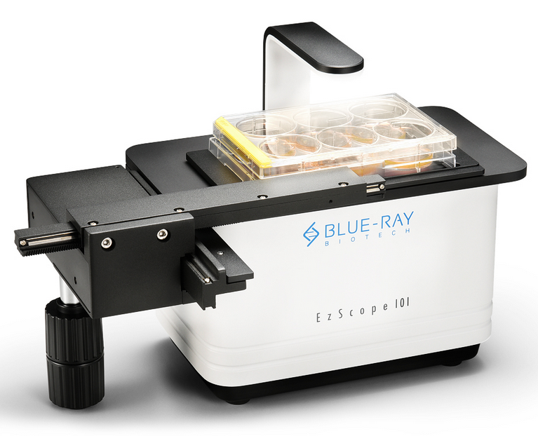
Live Cell Imaging EzScope
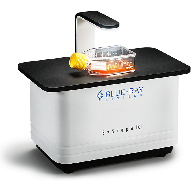

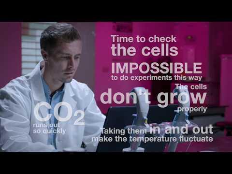
With EzScope 101 you can observe the cell behavior of your cultures without removing dishes or flasks from the incubator.
The cells can be viewed at any time as the software transfers the data live to your PC. EzScope 101 enables 24/7 measurements under precisely controlled conditions in a non-perturbing environment.
Specifications
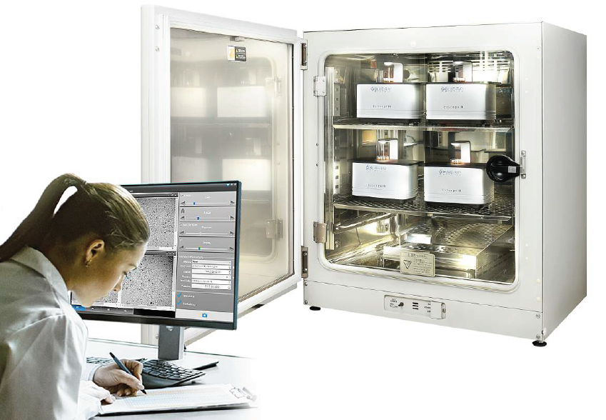
- For up to four samples in one incubator simultaneously (also CO2 incubator)
- Two lenses (easily interchangeable)
- Motorized focusing (control via PC, incubator does not have to be opened)
EzScope-101 - Live Cell Imaging System
Applications
- Cell growth and confluence
- Cell migration and wound healing
- Stem cell behaviors
- Cell death assays
- Spheroid development and behaviors
- Cultivation of yeast
Incubator Live View
Designed to be used inside the incubator, without the need to remove your cells from incubator to enhance culture quality control.
Minimizes Experimental Variations
Up to four units of EzScope can to be setup in the same incubator and controlled by one computer. This enables the monitoring of samples simultaneously, reduces errors caused by environment variations.
Exceptional Image Quality
Adopts high contract brightfield optical configuration, coupled with precise motorized focusing, and two interchangeable magnifying objective lenses.
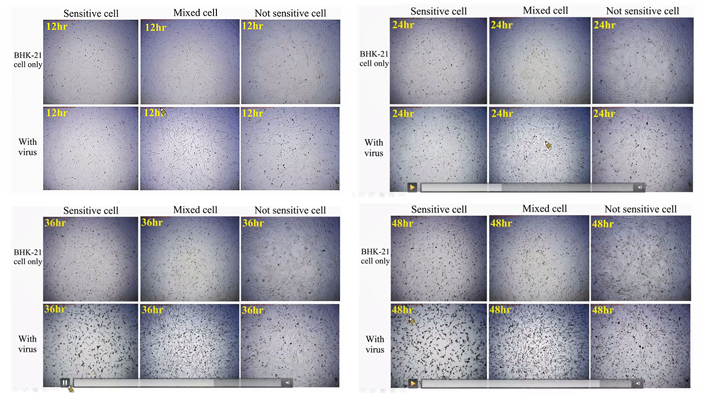
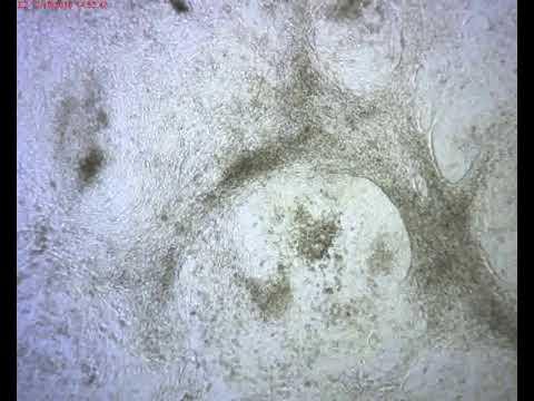
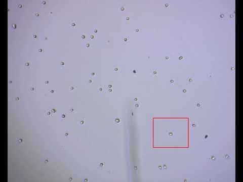
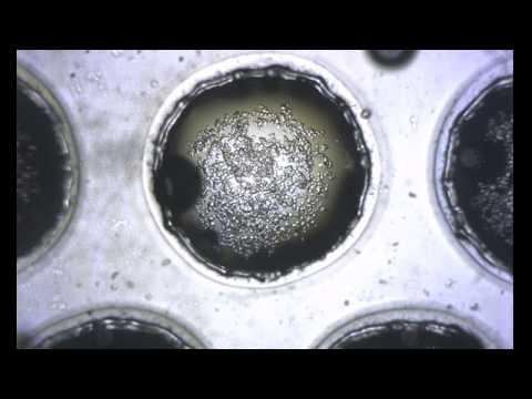
Image Examples:

Remote Monitoring of Experiment
Allows flexible remote monitoring the assay via Windows-based remote desktop software.
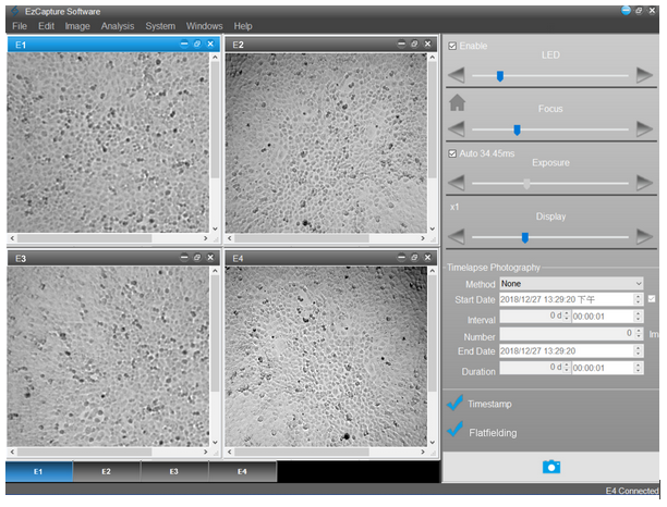
Easy Image Editor
Captures and edits images easily with EzCapture software:
- Live preview for up to 4 units of EzScope
- Flatfielding correction for even brightfield background
- Time-lapse video output
- Spatial calibration
- Measure and convergence analysis
Timelaps Imaging
| Order No. | 115.1900 |
| Optics | Brightfield (transmitted) with white LED |
| Objecitve Lens | 10x, 20x (optional) |
| Camera | 1.3 MP CMOS Sensor |
| Image Resolution | 1280 x 1024 pixels |
| Export Formats | Tiff (image), AVI (video) |
| Software | EzCapture with snapshot, time-lapse and confluence, etc |
| Field of View | 2.6 x 2.0 mm (10x objektive) |
| Resolution | 2 μm/pixel (10x objektive) 1μm/pixel (20x objektive) |
| Live View Frame Rate | Up to 8 frames/second |
| Focusing | Motorized |
| Stage | |
| Manual XY stage (optional) | SBS footprint |
| Labware Holders (optional) | 35 mm culture dish and slide, 60 mm culture dish and slide, T-25 culture flask, T-75 culture flask |
| General | |
| Computer Requirements | i3 CPU with 4 GB RAM, Windows 10 OS, i5 CPU with 8 GB RAM, Windows 10 OS for multiple units connection |
| Connectivity | USB 2.0/3.0, up to 4 units |
| Power Supply | Input: AC 100-240 V, 50/60 Hz, Output: DC 5 V, 2 A |
| Dimensions (W x D x H) | 225 x 131 x 205 mm |
| Weight | 2.0 kg |
| Operating Conditions | 0°C - 42°C |
| Relative Humidity | 5% - 95% non-condensing |
| Certifications | CE, RoHS |
Accessories (optional)
Manual XY stage (SBS Footprint)
Adapters for XY stage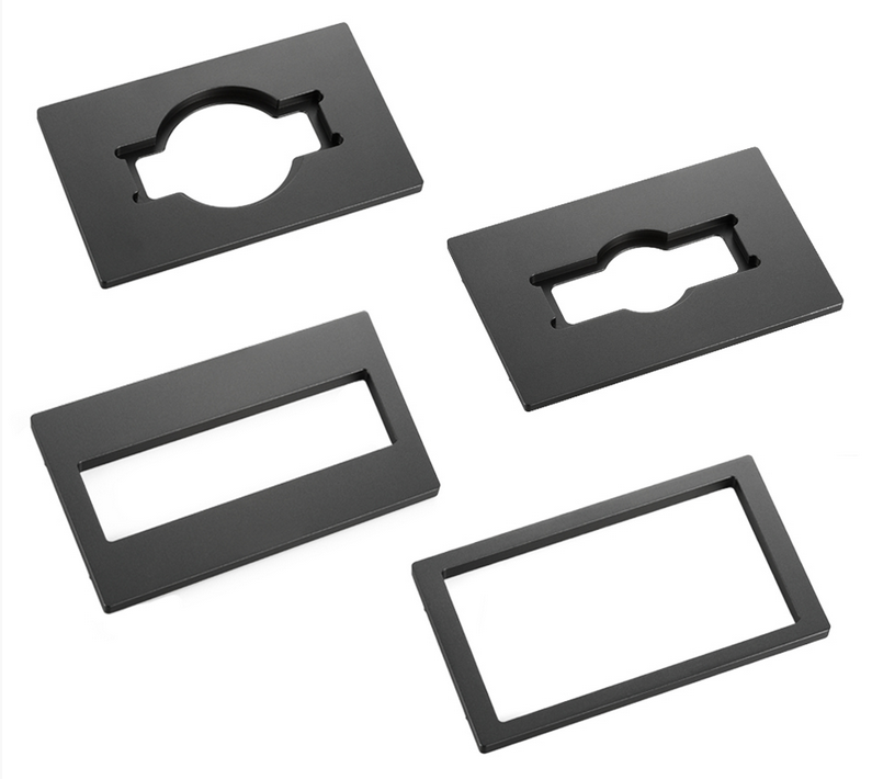
Which objective lenses are available for EzScope 101?
Standard 10x objective lens included; 20x objective lens also available for optional purchase. Users can switch lenses based on their applications.
What’s the field of view (FOV) range for each objective lens?
The FOV range of the 10X objective lens is 2.6 mm X 2.0 mm, and the FOV range for the 20X objective lens is 1.3 mm X 1.0 mm.
Will the high humidity environment of the incubator affect the device?
EzScopes has been rigorously tested and can endure such environment. However, in order to reduce additional corrosion caused to the device due to condensed water droplets, we strongly recommend users leave EzScope for 1-2 hours after placing it in the incubator and wait for the temperatures of the machine and incubator to balance, and then connect the power of the machine and place samples inside for observation. Doing so can also reduce the temperature changes when the cell sample comes in contact with the carrier of the machine.
If I clamp the signal cable and power cable on the incubator door, will it cause leakages and increase CO2 consumption?
There will be very little impact. Of course, the most perfect cable guiding method is through the dedicated equipment port for the incubator. But if your incubator does not have an equipment port, you can still clamp the signal cable and power cable between the door and the hermetic gasket. The hermetic gasket in the accessory pack of EzScope can be used when necessary to coat where the cable is clamped in order to ensure there is no leakage at all, and further protect the cable.
How should I clean and disinfect EzScope?
You can simply wipe with the regular 70% alcohol in laboratories. Please beware not to use too much disinfectant and prevent liquids from leaking inside the machine.
How should I clean the lenses?
You can first blow away the dust on the lens surface with a blower, and then drop 1-2 drops of 70% alcohol on the lens tissue and wipe the lens lightly. Wipe from the center of the lens outward in a spiral path, and then use a new lens tissue to wipe off the residual liquid.
Can I use EzCapture to measure the size of the cells in the photo?
Yes; EzCapture is equipped with measuring functions; please refer to the operation manual for details.
Will there be more cell image analysis functions for EzScope?
We’ve always insisted on continual improvement and providing more better products. We will strive to provide more analysis functions; you can update the software for free from the official website of Blue-Ray Biotech in the future. In addition, there are many 3rd party software systems available on the market that can also be used. For example:Fiji (https://imagej.net/Fiji) or CellProfiler (https://cellprofiler.org/).
An error message appeared while installing the drivers on the computer system; what should I do?
EzCapture only supports the Win 10 system. Please confirm the version of your computer system, install and execute the software on the correct system.
How can I access the EzScope’s picture from home?
EzCapture will save the captured pictures on the control computer connected to EzScope. There are also many third-party remote access software systems available for use (please refer to the operation manual for details); you can use these software systems to access the pictures on the computer remotely. Please note that operations can only be performed under environments with internet connections.
How can I remove blurred images from the time-lapse video?
When the conditions of the incubator components are poor or when there is a vibration source nearby, it may cause some or all of the images in the time-lapse series video to become blurry; please first eliminate the vibration source.
Small Tip: In time-lapse recorded videos, you can first use the built-in video editing functions to remove the blurry images before outputting the video.

Add-ons